Ginny competes in dog agility and can apply her chiropractic diagnosis and treatment based on how Benny uses his body when doing agility too – by being able to access her chiropractic services on a consistent basis Benny has made improvement in his stride and sharp turns needed on course.
Animals cannot seek medical attention on their own. To be seen they must do something that gets the attention of their owner/caretaker. As veterinarians sometimes, it can be difficult to ascertain where an animal is hurting. Thermographic imaging can help assist in these difficult cases. It can help detect anatomical areas that require diagnostic evaluation (manual evaluation, x-rays, ultrasound, MRI, CT).
This can also help determine if the area we are seeing clinical signs associated with are the original problem or compensation. Thermographic imaging can also help veterinarians monitor a patient’s response to the therapy we are using to be sure it is the right therapy for that pet. For dogs that participate in activities we can use the Thermographic imaging to help see problem areas before the pet even shows outward signs of the issue. The thermographic imaging can help with anxious or aggressive animals as it is noninvasive.
Thermographic does not depict temperature but measures the radiated energy from the target tissues. Different levels of this radiated energy are represented as different colors, this is referred to as a thermogram. The camera detects energy directly from the subject; therefore, there is no prejudice or outside influence on the results during an examination. Thermal detectors only measure energy entering the camera lens they emit nothing. Digital thermal imaging provides a representation of temperature difference. There are pattern formations (thermal gradients) associated with various areas of the body. It will not give you a diagnosis but allow you to focalize your other diagnostics or treatments. Physiologic activity will show with increased radiant areas and areas of neural irritation/dysfunction would show as less radiant. For each patient their own imaging serves are their own normal. So, we look for differences in opposites sides and confirm these findings by taking a second image.
The science behind this technology
The FDA fully recognized digital thermography as an adjunctive or supportive evaluation of the patient. There are at this time over 1,814 scientific papers listed under Digital Thermography on the U.S. National Library of Medicine’s website www.PubMed.gov. The electromagnetic spectrum includes the infrared portion which is outside what our eyes can see. All objects emit infrared radiation that the digital thermal equipment can record as an image. Temperature is the measure of the average kinetic vibrational energy of the molecules of that substance. Relative temperature measurement is calculated via the software within the digital imaging system. Thus, digital thermography is the collection and analysis of radiated electromagnetic energy in the infrared portion of the electromagnetic spectrum using a thermal imaging device. Different levels of this energy are displayed as different colors. All areas should be thermographically symmetrical. A difference in the symmetry of the thermal gradients indicates the need for further evaluation.
Example case
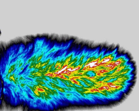
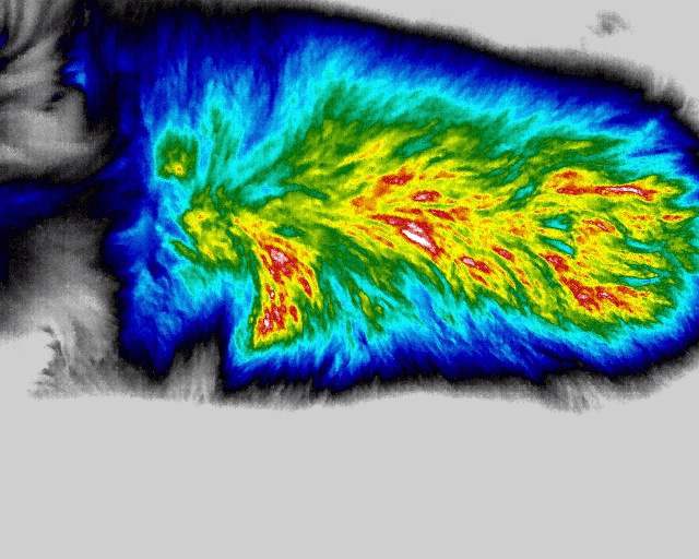
The picture of the dog at the top was just a little quiet the day it presented but nothing could be found by owner. No limping was present when patient was examined. As you can see there was a radiant area at the left shoulder. On further examination we found a trigger point in that area as well as areas that required spinal manipulation. Post treatment you can see the areas of radiant energy are dissipated at the left shoulder in the picture below.